Shoulder Muscles Diagram / Guide To Shoulder Anatomy. The extrinsic muscles of the shoulder include trapezius, latissimus dorsi, levator scapulae, rhomboid major. The shoulder muscles are associated with movements of the upper limb. If you know where muscles attach and how. Superficial layer with deltoid, trapezius, pectoralis major and. The primary function of the shoulder girdle is to give.
12 photos of the shoulder muscles and tendons diagram. We have five muscle diagrams of the shoulder. Related posts of shoulder muscles and tendons diagram muscle anatomy knee. Free access interactive and dynamic medical illustration of the shoulder's muscles : Initiates and assists deltoid in abduction of the arm and acts with rotator cuff muscles.
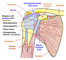
A Better Understanding Of Shoulder Health Girls Gone Strong Muscle Anatomy Shoulder Anatomy Shoulder Muscle Anatomy from i.pinimg.com They produce the characteristic shape of the shoulder, and can be divided into two groups The shoulder muscles bridge the transitions from the torso into the head/neck area and into the upper extremities of the arms and hands. The system used here groups the muscles based on their function and. Printable shoulder muscles diagrams to help you study the muscles structure in human's shoulder. Tutorials on the shoulder muscles (e.g rotator cuff muscles: Simple easy notes for quick revision for exams. There are three main muscles in your shoulder: Supraspinatus, infraspinatus, ters minor,.et), using interactive animations and labeled diagrams.
Learn vocabulary, terms and more with flashcards, games and other study tools.
The shoulder is not a single joint, but a complex arrangement of bones, ligaments, muscles, and tendons that is better called the shoulder girdle. Muscles of the shoulder can be subdivided into a variety of groups depending on origin, topography, function or innervation. Tutorials on the shoulder muscles (e.g rotator cuff muscles: Human muscles enable movement it is important to understand what they do in order to diagnose here we explain the major muscles of the human body. Human anatomy diagrams show internal. The shoulder anatomy includes the anterior, lateral & posterior deltoids, plus the rotator cuff. We have five muscle diagrams of the shoulder. Shoulder anatomy images shoulder muscle tissues anatomy actions diagram. The muscles in the shoulder aid in a wide range of movement and help protect and maintain the main shoulder this diagram with labels depicts and explains the details of shoulder muscles pictures. Related online courses on physioplus. The extrinsic muscles of the shoulder include trapezius, latissimus dorsi, levator scapulae, rhomboid major. The shoulder muscles include skeletal muscles that are attached to the head of the humerus which performs various direct and indirect functions of the shoulder joints. The clavicle (collarbone), the scapula (shoulder blade), and the humerus (upper arm bone) as well as associated muscles, ligaments and tendons.
The extrinsic muscles of the shoulder include trapezius, latissimus dorsi, levator scapulae, rhomboid major. In this video we'll explore the muscles and functions of the shoulder girdle (pectoral girdle). Learn vocabulary, terms and more with flashcards, games and other study tools. Muscles of the shoulder can be subdivided into a variety of groups depending on origin, topography, function or innervation. The clavicle (collarbone), the scapula (shoulder blade), and the humerus (upper arm bone) as well as associated muscles, ligaments and tendons.
Shoulder Wikipedia from upload.wikimedia.org Muscles of the shoulder are a group of muscles surrounding the shoulder joint, which move and provide support to the said joint. The muscles of the shoulder are associated with movements of the upper limb. The shoulder muscles produce the characteristic shape of the shoulder and can be classified into two. The extrinsic muscles of the shoulder include trapezius, latissimus dorsi, levator scapulae, rhomboid major. Free access interactive and dynamic medical illustration of the shoulder's muscles : 12 photos of the shoulder muscles and tendons diagram. They produce the characteristic shape of the shoulder, and can be divided into two groups Related online courses on physioplus.
Human anatomy diagrams show internal.
Superficial layer with deltoid, trapezius, pectoralis major and. Muscles of the shoulder can be subdivided into a variety of groups depending on origin, topography, function or innervation. Printable shoulder muscles diagrams to help you study the muscles structure in human's shoulder. In the above diagram, i added the latissimus dorsi muscle at 7. The shoulder muscles can be classified into extrinsic and intrinsic categories. If you know where muscles attach and how. The muscular system is responsible for movement in collaboration with the nervous system to form impulses for motion. The human shoulder is made up of three bones: The primary function of the shoulder girdle is to give. The shoulders are called the deltoid muscles or the deltoids. The shoulder muscles produce the characteristic shape of the shoulder and can be classified into two. Test your knowledge in our quiz about the shoulder muscles. Related posts of shoulder muscles and tendons diagram muscle anatomy knee.
This diagram depicts shoulder muscle diagram. Supraspinatus, infraspinatus, ters minor,.et), using interactive animations and labeled diagrams. The system used here groups the muscles based on their function and. This is a table of skeletal muscles of the human anatomy. Free access interactive and dynamic medical illustration of the shoulder's muscles :
Shoulder Muscles Anatomy And Functions Kenhub from thumbor.kenhub.com Learn vocabulary, terms and more with flashcards, games and other study tools. Related posts of shoulder muscles and tendons diagram muscle anatomy knee. In the above diagram, i added the latissimus dorsi muscle at 7. You'll need to build out all of these muscles if you want strong, balanced. Printable shoulder muscles diagrams to help you study the muscles structure in human's shoulder. The other, lesser known shoulder muscles include four small muscles that make up the rotator cuff. The extrinsic muscles of the shoulder include trapezius, latissimus dorsi, levator scapulae, rhomboid major. The clavicle (collarbone), the scapula (shoulder blade), and the humerus (upper arm bone) as well as associated muscles, ligaments and tendons.
Supraspinatus, infraspinatus, ters minor,.et), using interactive animations and labeled diagrams.
Learn vocabulary, terms and more with flashcards, games and other study tools. Human muscles enable movement it is important to understand what they do in order to diagnose here we explain the major muscles of the human body. Cuff muscles of the back shoulder muscles and chest human anatomy diagram the shoulder muscles bridge the transitions. Learn faster with interactive shoulder. Simple easy notes for quick revision for exams. The shoulder muscles are associated with movements of the upper limb. What's important to note here is that from. Human anatomy diagrams show internal. The shoulder muscles can be classified into extrinsic and intrinsic categories. Initiates and assists deltoid in abduction of the arm and acts with rotator cuff muscles. This diagram depicts shoulder muscle diagram. Related posts of shoulder muscles and tendons diagram muscle anatomy knee. The next life study seated female figure, shows the upper part of the the muscles of the back move the shoulder blade (scapula), upper arm (humerus), and back (vertebral.
Share :
Post a Comment
for "Shoulder Muscles Diagram / Guide To Shoulder Anatomy"


Post a Comment for "Shoulder Muscles Diagram / Guide To Shoulder Anatomy"